Follow Us:
Winner99E Static Image Particle Size Analyzer
Home  /  products  /  Particle Image Analyzer  /  Winner99E Static Image Particle Size Analyzer
  • Winner99E Static Image Particle Size Analyzer

  • Winner99E Static Image Particle Size Analyzer

Winner99E Static Image Particle Size Analyzer

Measurement Range: 1-6000μm

It is widely applicable to high-end application scenarios such as research institutes, university laboratories, and enterprise R&D centers and quality testing institutions.

Products Description

Winner99E Static Image Particle Size Analyzer

Winner99E is the latest generation of static particle image analysis equipment in China. It uses the latest image acquisition system and analysis software, perfectly combining computer imaging with particle size and shape analysis theory. While obtaining clear particle images, the particle size, sphericity, aspect ratio, bulkiness, surface ratio and other relevant particle size and shape characterization parameters are presented in the form of characteristic values and distribution, allowing users to understand the particles in detail. In addition, this instrument also has the symbols of the times of automation and intelligence, making its operation easier, analysis smarter, and results more stable. It is the preferred partner and right-hand man for particle size testing and particle shape analysis.

 Main Specifications


General
Microscopic System
Software Functions
Report Parameters
Model Name Winner99E
Measurement Range 1-6000μm
Maximum Resolution 2048*1536
Pixel Size 3.2*3.2μm
Segmentation Speed <1 second
Segmentation Success Rate >98%
Connection Port USB3.0
Eyepiece 10X wide field of view eyepiece
Objective Lense 4X, 10X, 40X, 100X (Oil) Long-Distance Chromatic Aberration Correcting (Plan) Objective Lens Group
Light Source Bottom-mounted translucent light source, halogen or LED light optional, brightness adjustable; top-mounted metallographic incident light source (with polarizer).
Platform Manual 3D mechanical stage, dimensions: 185mm x 140mm
Movement range: 50mm x 75mm, coarse and fine coaxial focusing
Fine adjustment increments: 2µm, with locking and limiting devices
Magnification 4-5000X
Image Acquisition Collect sample morphology images and save them in JPG and BMP formats.
Image Stitching Seamlessly stitching multiple images together allows for a greater number of particles in the test, improving its representativeness. Alternatively, individual images can be processed, saved, and then stitched together, further enhancing the accuracy of the results.
Automatic Processing Tools Twelve automatic processing tools, including automatic adaptive binarization, automatic noise removal, automatic removal of particles with incomplete boundaries, automatic filling of hollow areas in particles, automatic smoothing of particle edges, automatic elimination of particle adhesion, and automatic acquisition of particle size and shape parameters.
Scale After calibration with a national standard micrometer, each test only requires selecting the scale value corresponding to the objective lens to obtain the actual particle size.
Single-particle Data The image allows for direct display and analysis of 10 parameters for individual particles, including cross-sectional area, particle size, aspect ratio, and sphericity.
Task Management The task management mechanism is rigorous, enabling users to easily manage test data.
Feature Parameters Characteristic parameters of particle distribution such as D10/D50 (median diameter)/D90/D100
Frequency/Cumulative Data tables, graphs, and bar charts showing the frequency and cumulative distribution of particles by quantity, volume, and area.
Shape Parameters Ten commonly used data for characterizing particle shape, including aspect ratio, bulkiness, sphericity, surface area, specific surface area, and circumscribed rectangle parameter.
Number of Counts The number of particles tested can be obtained directly.
Thumbnails The report can display color or black-and-white (selectable) thumbnails of the samples.
Table Geader Specifications The report header allows you to enter information such as sample name, testing unit, and dispersion medium.
+ Advantages

(1) Intuitively reflect the particle morphology

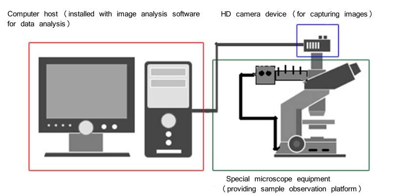
The surface morphology of the particles is directly reflected on the computer screen, so users can understand the morphology of the particles intultively and comprehensively.


(2) Perfectly stitch multiple images together

The particle images of different fields of view are stitched into one, so that more particles are involved in the analysis and the test results are more representative.


(3) Automatically segment of stuck particles

Adopt more advanced particle recognition algorithm to automatically segment various shapes of adherent particles, significantly improving segmentation accuracy, reducing human involvement and effectively shortening image processing time.


(4) Adaptive binarization function

The adaptive binarization function is used to ensure that the image binarization process is not affected by the shooting light, thus avoiding the loss of particle information due to uneven light. This lays the foundation for the accuracy of subsequent processing.


(5) Optical path design

The particle image analysis software can automatically process the tool set, integrate binarization, eliminate incomplete frequency particles at the boundary, etc. It only takes one key to process the particle mage and generate the analysis results. The operation is simple and the results are reliable.


(6) Freely switch particle size units

The ruler selection supports multiple length units, including nanometers, micrometers and millimeters. It is convenient for users to further process particle images obtained by electron microscopy and other methods.


(7) Quickly process particles with special shapes

A unique processing algorithm is used for spherical particles. Particles can be directly analyzed without processing the original image. Even if the particles are adhered or overlapped, the analysis result will not be affected. The analysis efficiency of spherical particles is improved.

+ Test Principle
Winner99E Static Image Particle Size Analyzer 
+ Application
Winner 99E static particle image analyzer used in cement, ceramics, pharmaceuticals, paints, dyes, pigments, fillers, chemical products, catalysts, coal, sediment, dust, flour, food additives, pesticides, explosives, graphite, photographic materials, fuel , metal and non-metal powder, calclum carbonate, kaolin and other powder industry.
Winner99E Static Image Particle Size Analyzer
+ Patent Technology

(1). Optical bench design is protected by patent No.- ZL 2014 2 0378380.8.

(2). Three dimensional-optical bench alignment system is protected by patent No.- ZL 2013 2 0835882.4.

(3). MIE scattering principle application patent No.- ZL 2013 2 0812021.4.

(4). Dry particle size analyzer full sealed sample cuvette application is protected by patent No.- ZL.2011 2 0267646.8.

(5). Dual laser beam orthogonal application is protected by patent No.-ZL 2007 2 0025702.0.

(6). Powder dispersion pump design application is protected by patent No.-ZL 2007 2 0018648.7.

(7). Wet circulation installation is protected by patent No.-ZL2010 2 0593526.2.

+Test Report
Winner99E Static Image Particle Size Analyzer
Contact Us
GET IN TOUCH

Copyright © Jinan Winner Particle Instrument Stock Co., Ltd. All Rights Reserved | sitemap  

ORDER NOW!